Customer service hotline:


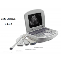


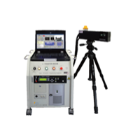
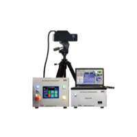
Technical parameters of LS-810 full digital ultrasonic diagnosis system
1. Display modes: B, 2B, 4B, B/M, M.
2. TGC control, frame correlation, gray scale transformation, edge enhancement, transverse filtering, dynamic filtering and other technologies, the whole image is clear, stable performance, high resolution.
3. Lateral resolution ≤3mm (depth ≤80), ≤4 mm (depth ≤130), axial resolution ≤2mm (depth ≤80), ≤3mm (depth ≤130), maximum detection depth ≥170mm, blind area ≤5.
4. Probe interface: 1.
5. Display: ≥10 inches
6. Image processing technology: controlled frame correlation, gamma correction, edge enhancement, image smoothing, image noise reduction, automatic gain adjustment, image up and down, left and right, (90º, 270º) flip;
7. Image amplification: stepless amplification; At the same time, it has real-time dynamic PIP picture-in-picture release function;
8. Image archiving, browsing, comparison, storage, printing, transmission functions; The machine can permanently store at least 100 images, storage picture slide mode full-screen browsing;
9. Support U disk, software can be automatically upgraded through U disk, and can store or read images on U disk;
10. Automatic measurement and calculation, ratio measurement, line narrowing ratio, surface narrowing ratio, Angle measurement; Puncture guidance, histogram, profile;
11. Image processing preset and one-click optimization function, doctors can pre-set more than 8 groups of parameters according to clinical needs;
12. Body mark: more than 40 kinds of body mark;
13. Continuous operation of the diagnostic system ≥8h
14. Puncture guide: with probe puncture guide device
15. Medical record information database system, patient information storage, retrieval, management functions;
16. Diagnostic measurement formula preset system, which can set different formulas according to different races;
17. Automatic measurement software for obstetrics, gynecology, small organs, heart, urology and other departments, automatic report generation system, connect laser printer can directly print A4 diagnostic report;
18. Configuration: one host, one convex array probe (abdomen), one linear array probe (small organ), and one cavity probe (negative probe).